What complications can occur after prostate cancer surgery?

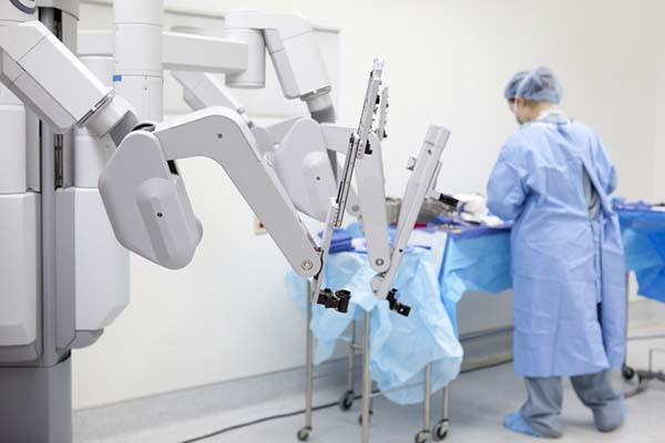
Earlier this year, US defense secretary Lloyd Austin was hospitalized for complications resulting from prostate cancer surgery. Details of his procedure, which was performed on December 22, were not fully disclosed. Press statements from the Pentagon indicated that Austin had undergone a minimally invasive prostatectomy, which is an operation to remove the prostate gland. Minimally invasive procedures are performed using robotic instruments passed through small “keyhole” incisions in the patient’s abdomen.
Just over a week later, Austin developed severe abdominal, hip, and leg pain. He was admitted to the intensive care unit at Walter Reed Hospital on January 2 for monitoring and further treatment. Doctors discovered that Austin had a urinary tract infection and fluid pooling in his abdomen that were impairing bowel functioning. The defense secretary was successfully treated, but then readmitted to the ICU on February 11 for what the Pentagon described as “an emergent bladder issue.” Two days after undergoing what was only described as a “non-surgical procedure performed under general anesthesia,” Austin was back at work. His cancer prognosis is said to be excellent.
Austin’s ordeal was covered extensively in the media. Although we cannot speculate about his specific case, to help our readers better understand the complications that might occur after a prostatectomy, I spoke with Dr. Boris Gershman, a urologist at Harvard-affiliated Beth Israel Deaconess Medical Center in Boston. Dr. Gershman is also a member of the advisory and editorial board for the Harvard Medical School Guide to Prostate Diseases.
How common are urinary tract infections after a prostatectomy?
Minimally invasive prostatectomy is generally well tolerated. In one study that examined complications among over 29,000 men who had the operation, the rate of urinary tract infections was only 2.1%. The risk of sepsis — a more serious condition that occurs if the body’s response to an infection damages other organs — is much lower than that.
How would a urinary tract infection occur?
Although urinary tract infections are rare after prostatectomy, bacteria can travel into the urinary system through a catheter. An important part of a prostatectomy involves connecting the urethra — which is a tube that carries urine out of the body — directly to the bladder after the prostate has been taken out. As a last step in that process, we pass a catheter [a soft silicone tube] through the urethra and into the bladder to promote healing. Infection risks are minimized by giving antibiotics both during surgery and then again just prior to removing the catheter one to two weeks after the operation.
How do you treat urinary infectious complications when they do happen?
It’s not unusual to find small amounts of bacteria in the urine whenever you use a catheter. Normally they don’t cause any symptoms, but if infectious complications do occur, then we’ll admit the patient to the hospital and treat with broad-spectrum antibiotics that treat many different kinds of bacteria at once. We’ll also obtain a urine culture to identify the bacterial species causing the infection. Based on culture results, we can switch to different antibiotics that attack those microbes specifically. The course of treatment generally lasts 10 to 14 days.
Lloyd Austin also had gastrointestinal complications. Why might that have occurred?
Although I cannot speculate about Austin’s specific case, in general gastrointestinal complications are very rare — affecting fewer than 2% of patients treated using robotic methods. However, a few different things can happen. For instance, the small intestine can “fall asleep” after surgery, meaning it temporarily stops moving food and wastes through the bowel.
This is called an ileus. It can be due to multiple reasons, including as a result of anesthetics or pain medications. An ileus generally resolves on its own if patients avoid food or water by mouth for several days. If it causes too much pressure in the bowel, then we “decompress” the stomach by removing accumulated fluids through a nasogastric tube, which is threaded into the stomach through the nose and throat.
Some patients develop a different sort of surgical complication called a small bowel obstruction. We treat these the same way: by withholding food and water by mouth and removing fluids with a nasogastric tube if necessary. If the blockages are caused by scar tissues, in rare cases this may require a second surgery to fix the obstructing scar tissue.
Fluids might also collect in the pelvis after lymph nodes are removed during surgery. What’s happening in these cases?
Pelvic lymph nodes that drain the prostate are commonly removed during prostatectomy to determine if there is any cancer spread to the lymph nodes. A possible risk from lymph node removal is that lymph fluid might leak out after the procedure and pool up in the pelvis. This is called a lymphocele. Most lymphoceles are asymptomatic, but infrequently they may become infected. When that happens, we treat with antibiotics, and we might drain the lymphocele using a percutaneous catheter [which is placed through the skin]. Fortunately, newer surgical techniques are helping to ensure that lymphoceles occur very rarely.
Are there individual factors that increase the risk of prostatectomy complications?
Certainly, patients can have risk factors for infection. Diabetes, for instance, can inhibit the immune system, especially when patients have poor glycemic or glucose control [a limited ability to maintain normal blood sugar levels]. If patients have autoimmune diseases, or if they’re taking immunosuppressive medications, they may also be at increased risk of infectious or wound healing complications with surgery, and in some cases, may instead be treated with radiation to avoid these risks.
Thanks for walking me through this complex topic! Any parting thoughts for our readers?
It’s important to discuss the potential risks of surgery with your doctor so you can be fully informed. That said, prostatectomy these days using the minimally invasive approach has a very favorable risk profile. The majority of patients do really well, and fortunately severe complications requiring hospital readmission are very rare.
About the Author

Charlie Schmidt, Editor, Harvard Medical School Annual Report on Prostate Diseases
Charlie Schmidt is an award-winning freelance science writer based in Portland, Maine. In addition to writing for Harvard Health Publishing, Charlie has written for Science magazine, the Journal of the National Cancer Institute, Environmental Health Perspectives, … See Full Bio View all posts by Charlie Schmidt
About the Reviewer

Marc B. Garnick, MD, Editor in Chief, Harvard Medical School Annual Report on Prostate Diseases; Editorial Advisory Board Member, Harvard Health Publishing
Dr. Marc B. Garnick is an internationally renowned expert in medical oncology and urologic cancer. A clinical professor of medicine at Harvard Medical School, he also maintains an active clinical practice at Beth Israel Deaconess Medical … See Full Bio View all posts by Marc B. Garnick, MD

What is a tongue-tie? What parents need to know

The tongue is secured to the front of the mouth partly by a band of tissue called the lingual frenulum. If the frenulum is short, it can restrict the movement of the tongue. This is commonly called a tongue-tie.
Children with a tongue-tie can’t stick their tongue out past their lower lip, or touch their tongue to the top of their upper teeth when their mouth is open. When they stick out their tongue, it looks notched or heart-shaped. Since babies don’t routinely stick out their tongues, a baby’s tongue may be tied if you can’t get a finger underneath the tongue.
How common are tongue-ties?
Tongue-ties are common. It’s hard to say exactly how common, as people define this condition differently. About 8% of babies under age one may have at least a mild tongue-tie.
Is it a problem if the tongue is tied?
This is really important: tongue-ties are not necessarily a problem. Many babies, children, and adults have tongue-ties that cause them no difficulties whatsoever.
There are two main ways that tongue-ties can cause problems:
- They can cause problems with breastfeeding by making it hard for some babies to latch on well to the mother’s nipple. This causes difficulty with feeding for the baby and sore nipples for the mother. It doesn’t happen to all babies with a tongue-tie; many of them can breastfeed successfully. Tongue-ties are not to blame for gassiness or fussiness in a breastfed baby who is gaining weight well. Babies with tongue-ties do not have problems with bottle-feeding.
- They can cause problems with speech. Some children with tongue-ties may have difficulty pronouncing certain sounds, such as t, d, z, s, th, n, and l. Tongue-ties do not cause speech delay.
What should you do if think your baby or child has a tongue-tie?
If you think that your newborn is not latching well because of a tongue-tie, talk to your doctor. There are many, many reasons why a baby might not latch onto the breast well. Your doctor should take a careful history of what has been going on, and do a careful examination of your baby to better understand the situation.
You should also have a visit with a lactation specialist to get help with breastfeeding — both because there are lots of reasons why babies have trouble with latching on, and also because many babies with a tongue-tie can nurse successfully with the right techniques and support.
Talk to your doctor if you think that a tongue-tie could be causing problems with how your child pronounces words. Many children just take some time to learn to pronounce certain sounds. It is also a good idea to have an evaluation by a speech therapist before concluding that a tongue-tie is the problem.
What can be done about a tongue-tie?
When necessary, a doctor can release a tongue-tie using a procedure called a frenotomy. A frenotomy can be done by simply snipping the frenulum, or it can be done with a laser.
However, nothing should be done about a tongue-tie that isn’t causing problems. While a frenotomy is a relatively minor procedure, complications such as bleeding, infection, or feeding difficulty sometimes occur. So it’s never a good idea to do it just to prevent problems in the future. The procedure should only be considered if the tongue-tie is clearly causing trouble.
It’s also important to know that clipping a tongue-tie doesn’t always solve the problem, especially with breastfeeding. Studies do not show a clear benefit for all babies or mothers. That’s why it’s important to work with a lactation expert before even considering a frenotomy.
If a newborn with a tongue-tie isn’t latching well despite strong support from a lactation expert, then a frenotomy should be considered, especially if the baby is not gaining weight. If it is done, it should be done early on and by someone with training and experience in the procedure.
What else should parents know about tongue-tie procedures?
Despite the fact that the evidence for the benefits of frenotomy is murky, many providers are quick to recommend them. If one is being recommended for your child, ask questions:
- Make sure you know exactly why it is being recommended.
- Ask whether there are any other options, including waiting.
- Talk to other health care providers on your child’s care team, or get a second opinion.
About the Author

Claire McCarthy, MD, Senior Faculty Editor, Harvard Health Publishing
Claire McCarthy, MD, is a primary care pediatrician at Boston Children’s Hospital, and an assistant professor of pediatrics at Harvard Medical School. In addition to being a senior faculty editor for Harvard Health Publishing, Dr. McCarthy … See Full Bio View all posts by Claire McCarthy, MD

Is chronic fatigue syndrome all in your brain?

Chronic fatigue syndrome (CFS) –– or myalgic encephalomyelitis/chronic fatigue syndrome (ME/CFS), to be specific –– is an illness defined by a group of symptoms. Yet medical science always seeks objective measures that go beyond the symptoms people report.
A new study from the National Institutes of Health (NIH) has performed more diverse and extensive biological measurements of people experiencing CFS than any previous research. Using immune testing, brain scans, and other tools, the researchers looked for abnormalities that might drive health complaints like crushing fatigue and brain fog. Let’s dig into what they found and what it means.
What was already known about chronic fatigue syndrome?
In people with chronic fatigue syndrome, there are underlying abnormalities in many parts of the body: The brain. The immune system. The way the body generates energy. Blood vessels. Even in the microbiome, the bacteria that live in the gut. These abnormalities have been reported in thousands of published studies over the past 40 years.
Who participated in the NIH study?
Published in February in Nature Communications, this small NIH study compared people who developed chronic fatigue syndrome after having some kind of infection with a healthy control group.
Those with CFS had been perfectly healthy before coming down with what seemed like just a simple “flu”: sore throat, coughing, aching muscles, and poor energy. However, unlike their experiences with past flulike illnesses, they did not recover. For years, they were left with debilitating fatigue, difficulty thinking, a flare-up of symptoms after exerting themselves physically or mentally, and other symptoms. Some were so debilitated that they were bedridden or homebound.
All the participants spent a week at the NIH, located outside of Washington, DC. Each day they received different tests. The extensive testing is the great strength of this latest study.
What are three important findings from the study?
The study had three key findings, including one important new discovery.
First, as was true in many previous studies, the NIH team found evidence of chronic activation of the immune system. It seemed as if the immune system was engaged in a long war against a foreign microbe — a war it could not completely win and therefore had to keep fighting.
Second, the study found that a part of the brain known to be important in perceiving fatigue and encouraging effort — the right temporal-parietal area — was not functioning normally. Normally, when healthy people are asked to exert themselves physically or mentally, that area of the brain lights up during an MRI. However, in the people with CFS it lit up only dimly when they were asked to exert themselves.
While earlier research had identified many other brain abnormalities, this one was new. And this particular change makes it more difficult for people with CFS to exert themselves physically or mentally, the team concluded. It makes any effort like trying to swim against a current.
Third, in the spinal fluid, levels of various brain chemicals called neurotransmitters and markers of inflammation differed in people with CFS compared with the healthy comparison group. The spinal fluid surrounds the brain and reflects the chemistry of the brain.
What else did study show?
There are some other interesting findings in this study. The team found significant differences in many biological measurements between men and women with chronic fatigue syndrome. This surely will lead to larger studies to verify these gender-based differences, and to determine what causes them.
There was no difference between people with CFS and the healthy comparison group in the frequency of psychiatric disorders — currently, or in the past. That is, the symptoms of the illness could not be attributed to psychological causes.
Is chronic fatigue syndrome all in the brain?
The NIH team concluded that chronic fatigue syndrome is primarily a disorder of the brain, perhaps brought on by chronic immune activation and changes in the gut microbiome. This is consistent with the results of many previous studies.
The growing recognition of abnormalities involving the brain, chronic activation (and exhaustion) of the immune system, and of alterations in the gut microbiome are transforming our conception of CFS –– at least when caused by a virus. And this could help inform potential treatments.
For example, the NIH team found that some immune system cells are exhausted by their chronic state of activation. Exhausted cells don’t do as good a job at eliminating infections. The NIH team suggests that a class of drugs called immune checkpoint inhibitors may help strengthen the exhausted cells.
What are the limitations of the study?
The number of people who were studied was small: 17 people with ME/CFS and 21 healthy people of the same age and sex, who served as a comparison group. Unfortunately, the study had to be stopped before it had enrolled more people, due to the COVID-19 pandemic.
That means that the study did not have a great deal of statistical power and could have failed to detect some abnormalities. That is the weakness of the study.
The bottom line
This latest study from the NIH joins thousands of previously published scientific studies over the past 40 years. Like previous research, it also finds that people with ME/CFS have measurable abnormalities of the brain, the immune system, energy metabolism, the blood vessels, and bacteria that live in the gut.
What causes all of these different abnormalities? Do they reinforce each other, producing spiraling cycles that lead to chronic illness? How do they lead to the debilitating symptoms of the illness? We don’t yet know. What we do know is that people are suffering and that this illness is afflicting millions of Americans. The only sure way to a cure is studies like this one that identify what is going wrong in the body. Targeting those changes can point the way to effective treatments.
About the Author

Anthony L. Komaroff, MD, Editor in Chief, Harvard Health Letter
Dr. Anthony L. Komaroff is the Steven P. Simcox/Patrick A. Clifford/James H. Higby Professor of Medicine at Harvard Medical School, senior physician at Brigham and Women’s Hospital in Boston, and editor in chief of the Harvard … See Full Bio View all posts by Anthony L. Komaroff, MD